Monday, August 26, 2013

On working in Food Service

As I've mentioned here before, I spent the last year and change working as a server at a small restaurant in my college town. I originally took this job thinking I would wind up having more time to blog, but that turned out not to be the case. In a given week I'd spend between 40-50 hours at the restaurant, mostly working double shifts. I'd show up at 10:30 am, have a break for about an hour and a half in the middle of the day, and then leave work anywhere between 9 and 10 pm. Between work and applying to graduate school, I had very little time and energy leftover to devote to my blog. However it all paid off because I'm starting graduate school and actually saved up some money to be able to go.

If I could do it again, I think I'd do everything the same way. Maybe I'd put my foot down a little harder about the number of hours I worked, but to be honest, I loved working in the restaurant. It had its ups and downs like any job, but at least it was never boring and I wasn't just sitting in front of a computer screen all day. I had fun, I met some great people, and learned so much about work and life. I also learned how little people understand the service industry. It may not have anything to do with science, but I wanted to write two lists. The first about what food service taught me, the second about what patrons should know about food service.

What Food Service taught me:

  1. Patience. Someone is angry with the way their food turned out and wants to spend 10 minutes (i.e. forever in restaurant time) telling you everything that's wrong with it. A customer thinks he's ready to order but spends another five minutes hemming and hawing over the menu while you're trapped there. Just breathe, keep your cool, and wait and listen. The world will keep spinning.
  2. Multi-tasking. There's an ass in every chair and everyone needs something. Run table 1's food, on your way back grab table 5's check and ask how table 6 is doing. Run table 5's check and then stop by table 8 on the way back to take their order. Boom. Efficiency.
  3. Kill 'em with kindness. From the moment table 6 walked in you could see they were trouble. Maybe they were fighting in the car on the way to the restaurant. Maybe they've had a bad day and want to take it out on someone. Either way, if you walk up to them with a big friendly smile and a helpful disposition, you're going to save yourself a lot of grief, and if you're lucky they'll leave happier than when they came in.
  4. The squeaky wheel gets the grease. Customers who are vocal with their needs, comments, and complaints will get more focused service. HOWEVER. Customers who are vocal with their needs comments and complaints and are POLITE, PATIENT, AND KIND to their server get the best service. More on that later.
  5. Attitude is everything. The best attitude is positive, self-confident, but always willing to learn. The worst attitude is to think you don't need to learn anything new.
  6. Taking criticism is an important life skill. When taking criticism about yourself it's important to a. lower your defenses, and b. listen. When someone is telling you that you did something wrong, it's easy to hide behind your intentions. However intentions don't affect the world, actions do. So when someone is telling you how to do something better, even if they're yelling it at you in frustration, listen.
  7. Don't take everything personally. When you wait tables in a college town, you get a lot of bad tips, even when your service is great. When you wait tables anywhere, you get a lot of rude customers. People don't always say please, thank you, or even treat you like a human being when you're in the service industry. It doesn't take long for most servers to learn to not sweat the small stuff.
Things customers should consider when eating out:
  1. The minimum wage for servers in most of the US is $2.13 per hour. Servers literally make their living off of your tips, so don't be stingy. Even if your service was terrible you should at least tip 15%. Most servers aren't hired without experience and are therefore at least competent at their jobs, and chances are if they messed up it was just a goof or it wasn't even their fault (something went wrong in the kitchen or elsewhere.) After all, we all goof up at our jobs, but few of our jobs will pay us less when we make a mistake or two.
  2. Servers do not keep every penny of their tips. Most restaurants have a lot of support staff. Bussers, food runners, hosts, bartenders etc. that help the servers during the rush. Most restaurants also have a system where the servers must give away a portion of their tips to the support staff every night. This can mean that the server ends up walking home with only 60-70% of their tips. Moreover, tip-outs are usually calculated based off the server's sales, not their tips. So if you don't tip at all, then the 2% of your bill that goes to support staff that would normally come out of your tip comes out of the server's pocket instead. In other words, if you don't tip then your server essentially has to pay out the support staff themselves, and your server basically just paid for you to eat at their table.
  3. If you are vegan, gluten-free, or allergic to anything, tell your server BEFORE you order anything, and be honest. I can't tell you how many times I had a table order a vegetarian appetizer only to ask me, as they were eating their app, what was vegan on the menu. That puts me in an awkward spot. Do I tell them that the appetizer they're raving about has egg in it? Do I let it slide? I can't read your mind, you have to tell me before you order that you're vegan. When it comes to allergies, it pays to be honest. It's astonishing how many times people will insist they have allergies to something when they just don't like it or are avoiding it for diet reasons (ahem, gluten.) Just be honest. Servers actually want to make you happy. It's our job. If you want to avoid the gluten in the breading on the chicken, fine. But don't tell me you're severely gluten intolerant and then ask me for soy sauce (which is full of gluten.)
  4. If you have any time constraints, tell the host when you arrive. Then tell your server when you sit down. Five minutes before you have to catch your movie is not the time to tell me that you need your to-go boxes and check NOW. However, if you tell me ahead of time, I can be honest with you about what will come out quickest from the kitchen, and I can bring your check out with your food. Similarly, if you are in a rush, do not expect your ticket to be jumped to the front of the line. That is not fair to the other customers and you are not your server's one and only priority, either. If you are in a huge rush then consider fast food.
  5. If, for whatever reason, you have a problem with your food, let me know immediately. This is where the squeaky wheel gets the grease. If you tell me at the end of your meal that you hated your food, well there's not much I can do about it then. But if you just had a couple bites and it's too spicy, not spicy enough, too cold, or you just don't care for it, let me know as soon as you can. But I cannot stress enough how important it is to be polite and patient when alerting your server to any issues with your food. If you simply say "excuse me, but my food is cold, could you please heat it up for me?" Or, "I just don't care for dish, would it be possible for me to order something else?" you are going to get wayyyyyyyyyyyy better service than if you say "this is disgusting" with a big old frown. As servers, we get it, you come in with an expectation and when those expectations are not met, it's frustrating. But it's our job to make you happy, and we want you to be happy so help us to help you. Be polite, be understanding that if you order something else it might take a little bit for it to come out. My mother always said, "you catch more flies with honey than with vinegar."
  6. Whenever you eat out, take a look around the restaurant. Are they really busy? How many employees can you spot walking around? Knowing these questions can really help. On occasion I worked solo shifts at my restaurant. I was the only server, there was no host or busser. It amazed me how little people were aware of that and thus expected the service to be perfect. If you see 10 other tables and only one person working, then you can expect that your service is going to be a little slower. Restaurants are run by humans, after all.

Monday, August 12, 2013

Megalo-debacle: Did Discovery Really Commit a Faux-Pas?

For over two decades, Shark Week on Discovery Channel has been raising awareness of one of the ocean's most mysterious and powerful predators. Discovery originally started Shark Week with the purpose to dispel myths about the dangers of sharks, and to heighten the public's respect for the creatures. However, this year, many fans have felt outraged that Discovery may be straying further away from the original purpose of Shark Week. This year, Discovery unveiled the faux-documentary, Megalodon: The Monster Shark That Lives.

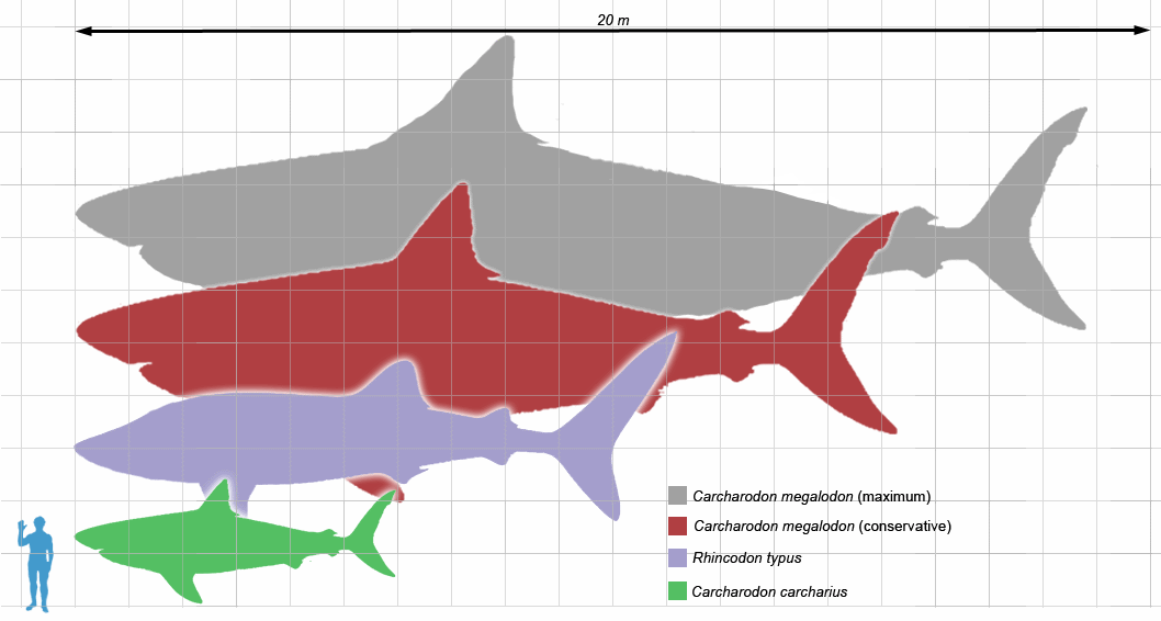
Megalodon, for the record, are definitely, absolutely extinct. They were super-sized sharks that once roamed the oceans some 2 million years ago.

Relative size of Megalodon (red and grey) vs. human. Source.

The Discovery special, on the other hand, suggested an alternative. Megalodon still roams the oceans, somewhere off the coast of South Africa. The documentary looked and seemed like any other documentary about real life events (however fantastic.) It convinced 70% of viewers that Megalodon could still live today. However, it was all fake. If you blinked you may have missed the disclaimers posted in small print:
"None of the institutions or agencies that appear in the film are affiliated with it in any way, nor have approved its contents."
"Though certain events and characters in this film have been dramatized, sightings of [the Megaladon,] 'Submarine' continue to this day."
"Megalodon was a real shark. Legends of giant sharks persist all over the world. There is still debate about what they may be."
These disclaimers appeared and disappeared quickly. Even if you had time to read them, they were still vague and beat around the bush. Nowhere do any of them directly say, "none of what you are viewing is based in fact."

It didn't take long for the blogosphere to ignite in outrage over the "documentary." Actor Wil Wheaton demanded Discovery apologize for misleading their audience. Popular science communicator, Christie Wilcox, wrote an open letter expressing her disappointment and anger with the direction Discovery has chosen to take with this year's Shark Week. Fans and scientists took to twitter to express their frustration. However, Discovery has stood by its documentary. Shark Week executive producer, Michael Sorensen, released this statement:
With a whole week of Shark Week Programming ahead of us, we wanted to explore the possibilities of Megalodon. It's one of the most debated shark discussions of all time, "can Megalodon exist today?" It's the ultimate Shark Week Fantasy. The stories have been out there for years and with 95 percent of the ocean unexplored, who really knows?
This statement is even more misleading. Asking "can Megalodon exist?" is not the same as asking, "does Megalodon exist?" which was the question the documentary was really asking.

When I first heard that the documentary was fake, I posted a link to Christie Wilcox's open letter on my facebook. It got several shares and comments from my friends who were as upset and disappointed as I was. Eventually my sister chimed in with a point that stopped me dead in my self-righteous tracks.
Maybe I'm missing something because I haven't seen [the show] but I'm not sure why it's generating this level of outrage. Annoyance, sure. Disappointment, totally fine. But that article is way over the top. They made a fake documentary and weren't so forthcoming with the "fake" bit (intentionally, I'm sure). They had 70% of viewers going for a minute there. Seems like that was probably the point right? They probably counted on the outrage from the science community to make their disclaimer for them. Success on all counts! Plus anyone who wasn't already aware now knows Shark Week has kicked off. I think expectations that Discovery is anything but a TV channel with a marketing plan are kind of off.
I think my sister, Laura, makes a lot of good points here and raises many important questions. First off: what responsibility does a TV channel have to present facts? They made the disclaimers, however vague and however quickly. And, as Laura pointed out, if there was anyone who missed the disclaimers, they certainly know now that the documentary was fake. Do we really have the right to be outraged? Are we, the "science community," just personally offended that Shark Week no longer meets our standards for good educational television? Or does Discovery have an obligation to uphold the original purpose and message of Shark Week from 26 years ago? I want to hear your thoughts. Discuss!

Monday, June 3, 2013

Getting Sprung: The Biological Underpinnings of Spring Fever

This article is being published here with permission from The Synapse. It originally appeared in the Spring 2013 edition of The Synapse at Oberlin College.


Before I was a student at Oberlin, the phrase “Spring Fever” meant little to me. However, once I matriculated the seasons grew more palpable. Perhaps it was the daily hikes around campus, but something about the air seemed to penetrate deeper into my skin. I felt especially vulnerable to the mood swings of Ohioan weather. I dealt with winter by resigning to it.

On the auspicious day when the clouds surrendered to sunshine and warmth, I was thrust out of my hibernation by the delicate savor of flowers and fresh grass. The sun filled me with a restless energy that invited me to skip class, sit out on North Quad, and look for four-leaf clovers with a friend. I partied later into the night and struggled to fall asleep as the birds chirped in the early morning. Over time, it became clear to me that many students are stricken with this same “fever” come spring. A particularly bright-eyed friend became especially reanimated in spring, proclaiming he was “solar powered.”

Talk to any Obie long enough and eventually you will learn the unique way that the seasonal changes in sunshine and warmth affect them. Their explanations range from the transformation of the monotonous winter grey into bright blue skies, longer days, warmer air, and a pleasant scent of renewal. But is there a more deep-seated biological rationale for such changes in mood and behavior? Is Spring Fever merely a social construct or is it an artifact of evolution?

“I would not be surprised if there was a biological imperative to go out and have fun in the spring,” muses Zachary Weil, an assistant professor at the Wexner Medical Center of Ohio State University. Weil, who studies seasonal changes in behavior and physiology in animals, speculates that the drive to get vitamin D from sunlight has something to do with Spring Fever. When the ultraviolet wavelengths in sunlight strike the skin, they stimulate light-reactive chemicals to synthesize vitamin D. The increased production of vitamin D in sunnier months may improve both physical and emotional health.

Overall, the scientific literature on Spring Fever is sparse. Just as it is impossible to appreciate light without darkness, scientists find it useful to study what drags us down in the winter, and to assume that the alleviation of those factors causes us to bounce back in the spring.

Melatonin, affectionately known as the hormone of darkness, is associated with seasonal changes in mood, behavior, and health. The pineal gland in the brain modulates the production of melatonin based on light levels. When light enters the eye, it stimulates neurons that connect to hypothalamus which tells the pineal gland to stop producing melatonin. However, if the pineal gland remained in darkness, it would modulate melatonin cyclically, approximately 10-hours-on, 14-hours-off. According to Weil, the pineal gland can sometimes “think” it is in darkness during the day.

“We're not aware of this consciously—because our eyes adjust so quickly—but the lights inside our offices and homes are orders of magnitude dimmer than the lights outside.” Sunlight, says Weil, is around 10,000 times brighter than incandescent and fluorescent bulbs. “People in northern climates that might go to work before the sun comes up and leave work after the sun goes down may never be exposed to the level of sunlight that's necessary to turn down our melatonin production.” Fathom the brain as an ancient machine responding to archaic devices such as the eyes and ears and it is conceivable that the brain may interpret this situation as perpetual darkness.

Conversely, once the days begin to lengthen and people are exposed to more morning sunlight, the brain produces melatonin for shorter intervals. The difference in melatonin production in the winter versus the spring is the predominant rationale for the prevalence of winter depression, or Seasonal Affective Disorder (SAD) in northern latitudes. Melatonin's effect on mood and behavior is complicated, though. Even though longer periods of melatonin production have been associated with SAD, melatonin can also be used as a treatment for people suffering from winter depression. Experimental therapies have shown that depending on when the dose is given, in conjunction with the patient's natural sleep-wake cycle, melatonin can actually help regulate the circadian rhythm and relieve depression. In general, melatonin production that begins in the evening and stops in the early morning helps most people combat the winter doldrums, which mimics a springtime daylight cycle.

For Obies though, nothing competes with actual sunshine and warm air. Fourth-year Nicole's* fondest spring memory happened in the last few days of her first year. Having just pulled three consecutive all-nighters to finish a paper, she shifted her focus to an attractive classmate. “I remember running into him at Stevie, [the school dining hall] but even inside Stevie it smelled like spring.” Emboldened by the triumph of having just completed her freshman year, she decided to catch up with him later that night at a party. “I remember walking back home with him, and I don't know, there was just something in the air.”

Things fizzled out between Nicole and her spring fling, but her experience remains an idyllic memory of springtime in Oberlin. “I was totally giddy and euphoric. It was a very spring collegiate freshman year experience.” Nicole's experience is one of many similar ones from several students I interviewed about their experiences of springtime in Oberlin. Some ancient vestige of biology springs from the increase in vitamin D, the decrease in melatonin, mixed with end-of-the-year excitement to foster delight among the students.

*Name has been changed to protect the student's privacy.

Monday, April 29, 2013

MDMA, "Drugs Live," and a life update.

It's been a while.

Last summer, I became inspired to write an article about the potential benefits of the club drug, MDMA, otherwise known as Ecstasy or Molly. The blog post got turned into an article for my alma mater's science magazine, The Synapse, and was published a few months ago. With permission, I am cross-posting it here.

A quick life update for anyone who is interested will be at the bottom of this post.


In Europe and the United States, the drug known as ecstasy is the magic bullet that loosens the inhibitions of many dancers at all-night dance parties known as “raves.” Ecstasy—or “Molly” in its allegedly purer form—is a psychedelic drug with a signature high that produces an intense sensory experience coupled with feelings of euphoria and closeness with others. However, ecstasy's tendency to raise body temperatures and dehydrate users in over-packed, sweltering clubs creates a dangerous situation that troubles parents and politicians alike.

Though ecstasy has been the culprit behind some tragic deaths since its popularization in the mid-eighties, death and injury from ecstasy are relatively rare compared to other drugs scheduled I or II by the DEA. The number of emergency room visits per year resulting from the use of ecstasy are tens of thousands fewer than those due to the use of cocaine, heroin, and marijuana. Every year, the number of deaths from ecstasy are miniscule relative to tobacco- and alcohol-related deaths. Some experts believe that MDMA, the primary chemical constituent behind the ecstasy high, isn't the real danger of ecstasy. Instead, they believe that it is the crowded, hot dance floors combined with the effects of the many other substances ecstasy is famously adulterated with that puts users in danger.

Despite the risks, MDMA's signature high has not only beguiled party-goers. The drug has also intrigued many scientists with its potential therapeutic benefits. In hushed sessions behind closed doors, therapists in the seventies and eighties began to explore the drug's ability to release patients from painful emotions attached to traumatic experiences and to strengthen the therapist-patient alliance. Although the anecdotal evidence was in favor of MDMA as a therapeutic drug, no placebo-controlled clinical trials had been performed by 1985, which was when MDMA was on the table for scheduling by the DEA. Due to the lack of clinical data, and MDMA's perceived dangers in the club scene, the drug was labeled as Schedule 1: a harmful drug with no medical benefits. All research on the therapeutic effects of ecstasy were halted for the next two and a half decades.

Nevertheless, MDMA has only become more ubiquitous among young people since 1985, and the cries to research the actual effects of MDMA on the human body have gradually swelled to a dull roar. However, the US and British governments have little to no precedent for funding studies on MDMA in humans. In 2010, Channel 4 in London endowed Professors David Nutt and Val Curran with funding to begin the first fMRI study of MDMA's effects in the human brain. The results of the study were broadcast live in a TV special called Drugs Live: The Ecstasy Trial late last September.

In the trials, conducted in September 2011, 25 volunteers came in for testing twice. Each time a volunteer came in, they received either an 83 milligram dose of MDMA or a placebo. The study was double-blind, so neither the administrators nor the subjects knew which drug they were getting. 30 minutes after they took the pill, the volunteers entered a fMRI scanner, where they were monitored for 90 minutes while answering questions about their subjective experiences. Throughout this process, volunteers were also asked to recall positive and negative memories from their lives. After the volunteers came out of the scanner, they performed a task in which they rated the trustworthiness of various faces, testing their feelings of closeness with others.

Drugs Live features the experiences of five volunteers: an ordained priest, an ex-soldier, a journalist, an actor, and a former member of Parliament. The show itself consists of clips of the five volunteers' trials on ecstasy, interviews with them and members of the audience, a debate between David Nutt and his loudest dissenter, Andrew Parrott, short videos of recreational MDMA users out dancing or just enjoying a night in with friends, and a video of an illegal therapy session with MDMA. The program is punctuated by fleeting explanations of the results of the study, described by Nutt and host Jon Snow with the aid of a giant, plastic brain with flashing lights indicative of the various structures within.

The first major discovery presented in episode 1 is MDMA's effects on the neural circuit between the posterior cingulate cortex and the prefrontal cortex. This circuit is known to become overactive in people suffering from anxiety disorders and depression, and is believed to lead to the excessive rumination characteristic of mood disorders. Normally, the two nuclei fire in sync with one another, but MDMA releases a deluge of serotonin and causes the two nuclei to start firing out of line, hushing the circuit between them and alleviating anxiety, which leads to the characteristic euphoria of the drug.

After a clip showing a therapy session, in which a woman talks through her feelings towards her recently deceased, abusive father, Nutt walks toward the giant brain to explain how MDMA is helping this woman work through her traumatic memories. When a person recalls a traumatic memory, there is an activation of the amygdala and the prefrontal cortical region. According to Nutt, the prefrontal cortical region modulates the emotions associated with a particular memory, and MDMA works to dampen the firing of that region. Without the chatter of the emotional overlay, patients are better equipped to engage with and process those memories, making MDMA a particularly exciting potential therapy for post traumatic stress disorder.

Though the program opened with a claim of being politics-free, “unvarnished science,” many viewers felt that Drugs Live was more of a “pro-drugs” circus than a lucid exposition of the science behind MDMA. Indeed, there was little discussion of the negative aspects of MDMA, including the “Tuesday blues” experienced by one volunteer in the study and the overall negative experience the ex-soldier had during the trials. There was also little air time for the debate between Nutt, Curran, and Parrott, which some viewers with a scientific background were particularly interested in.

Perhaps the positive bias in Drugs Live is unsurprising to those who know David Nutt for famously suggesting that drugs like cannabis, ecstasy, and LSD were less dangerous than alcohol and tobacco while on the Advisory Council on the Misuse of Drugs. His comments were largely responsible for his subsequent firing from the Council.

Regardless of Nutt's personal opinions on illicit substances, the study performed in Drugs Live is still an exciting contribution to a sparse body of research on the effects of MDMA in humans. Many other studies on MDMA are funded by government agencies such as the National Institute on Drug Abuse (NIDA). These studies tend to be performed on animals with the intention of finding the harmful neurological effects of MDMA. Such studies have shown significant depletion of neurons that produce serotonin that lasts for years after a single round of MDMA administration. However, these single doses are given intravenously over the course of three or four days. Critics say such dosages are not representative of how the drug would be administered in therapy (a low dose taken orally once or twice in a patient's lifetime), nor do they reflect how most recreational users take ecstasy (taken orally once or twice a month.)

Studies on humans are often relegated to surveying cognitive faculties in recreational users of ecstasy, using hair and urine drug tests to determine what sorts of drugs subjects have been taking. There are many criticisms of these studies as well, many citing poor control for subjects who have taken ecstasy in combination with other drugs or alcohol. However, these studies still show many deficits in memory, as well as higher levels of anxiety in current and former ecstasy users. Theseresults foster skepticism for the therapeutic benefits of MDMA.

A viewer watching Drugs Live likely wouldn't catch the troublesome findings of such research on MDMA just from watching the few short minutes where Andrew Parrott attempted to explain it. This was one of many issues that commenters brought up about Drugs Live after it aired. Some criticized Channel 4 for not giving enough credit to its viewers for being able to hold their attention on scientific facts for more than a few seconds. It seemed as though the content of Drugs Live wasn't much different from that of ecstasy found on the street: very little though potent science adulterated with an abundance of questionable filler.


Life update: For anyone who is wondering what I've been up to this past year, I'm still writing! I have also been working a lot waiting tables to save up for graduate school. I will be beginning the science communication graduate program at University of California Santa Cruz in the fall. In addition, I have been writing for The Synapse, and I will be posting my newest article once it comes out in print!

Wednesday, June 13, 2012

The Unsung Scientist, Louis-Antoine Ranvier

To many who read this blog, Notes of Ranvier is a title that probably evokes no thoughts of science or history. There is a backstory to the name, however, and a reason why I chose it as the title.

Notes of Ranvier is meant to be a play on words referring to the nodes of Ranvier, anatomical structures in certain types of neurons that have a myelin sheath. Every neuron has a long projection called an axon that transmits electrical signals to other neurons. Around the axons of some neurons is the myelin sheath, a fatty tissue that insulates the axon like plastic around a copper wire. Electricity can't travel though myelin, so there are even gaps between the sheath where the neuron is exposed and electrical currents can be propagated down the axon. These gaps were discovered by French scientist, Louis-Antoine Ranvier (pronounced rahn-vee-yeh), and thus bear his name as Ranvier's nodes or the nodes of Ranvier.

When you learn about Ranvier's nodes in class, not a lot of attention is paid to how they were discovered or why they have Ranvier's name instead of some other scientist. The treatment of the subject is far more along the lines of, "these exist, this is what they do, moving on." But the question still gnaws, who was Ranvier? How did he find these nodes, and how did he figure out what they do?

Ranvier was a histologist in 19th century France. Histology is the study of organic tissues, employing techniques such as staining and preserving tissues and examining them under a microscope to better understand their anatomy and physiology. When you looked at slides of cells dividing in high school biology, you were performing histology.

While histology and microscopy are so common today that even small children learn how to use microscopes in class, the microscope was renounced by many French scientists and doctors in the 19th century. The dismissal of the microscope was largely related to the dismissal of the cell. Even as late as the 1800's, the theory of the cell being the building block for all living things was still pretty far-fetched to many scholars. This idea known as cell theory, was hotly debated for centuries. But in the latter half of the tumult was Ranvier holed away in his laboratory, with his icepick of a microscope, diligently scratching and scraping on the surface of physiology.

From the time Ranvier was born in 1835 to the peak of his career, histology began to make a sea change in the eyes of French scholars. Histologists were developing newer and more advanced techniques for preserving and staining samples. Ranvier saw the merits of histology for studying anatomy and physiology. In particular, he valued the scrupulousness involved in preparing tissues for histological examination which allowed him to reveal the nodes between myelin sheaths and determine their function.


For a long time it was known that myelin sheaths existed and that they were made up of fatty cells. The cells were and still are called Schwann, after the scientist who identified them. Ranvier wanted to better understand how cells insulated by myelin sheaths were able to exchange nutrients (such as oxygen and ions) with the blood since carmine tissue stains demonstrated that such nutrients could not penetrate through Schwann cells. Closer examination with different chemicals and a more exacting technique revealed the nodes pictured below.
Ranvier's Nodes (e). Image from Barbara, J (2007). Originally from Ranvier, 1878.
Ranvier wasn't finished there. He still wanted to know whether the nodes really were the site for nutrient exchange between the neuron and the capillary. Around the neurons in your body is a small sheet of connective tissue that protects them from mechanical damage. Ranvier destroyed this tissue, and then poured water onto the exposed nerves of living animals. This caused the Schwann cells to swell and expand to cover the nodes. The result? A loss of neuron function and paralysis. Ranvier correctly deduced that the nodes were important for the conduction of signals through neurons.

Ranvier went on to develop some of the first legitimate theories on nerve cell degeneration with his experiments on myelin. Meanwhile, furious debate raged on about the nature of cells and their contributions to the function of the human body. Ranvier, all the while, was only interested in facts and improving his histological techniques. His mentality led to many great discoveries in his life, some of which contributed to our modern understanding of neurophysiology.

He was a man truly deserving of respect and admiration, and his approach to his work should be an inspiration to scientists and science writers alike.

Sources:
Bracegirdle, B. The History of Histology: A Review of Sources. (1977) Hist. Sci., 15:77-101

Barbara, J. Louis Ranvier (1835-1922): The Contribution of Microscopy to Physiology and the Renewal of French General Anatomy. (2007). Journal of the History of Neurosciences, 16:413-431.

Tuesday, May 15, 2012

Ranvier Returns

Hey everyone! I just wanted to write this to explain my absence.

In the last few weeks I left my job at the lab and decided to move back home to Ohio. I did this because I wanted more time to focus on what I really want to do, which is to write about science. The first step in that process, after the move, was to attend the science writer's workshop in Santa Fe, NM. I learned a lot there and came home feeling renewed and inspired. If you are reading this and are interested in getting into science writing yourself, I highly recommend you click that link above and apply to go to the workshop next year.

Over the next year I would like to write a lot more, but hopefully write for publications as well as here on this blog. So I'll be coming back with actual stories and new posts soon! I'm hoping to revise some of my older posts to make them better, too. If you want to stay up to date with me, follow me on twitter @NotesOfRanvier. I'll also be making a page on Facebook soon, too. So be on the look out!

Here are some photos from Santa Fe:


At the School for Advanced Research 
Prickly Pear Margaritas -- Yum!
I'm not really sure who this lady is or what she was doing but she looked cool so I took a picture.


More Notes of Ranvier soon.

Tuesday, March 13, 2012

Salty Penguins Filter Salt Out Their Nose

The Venture Brothers season 1, episode 5, via [adult swim]

Why yes, penguins do have an organ that converts sea water into fresh water! Except it's not an organ, it's a gland. And it doesn't directly convert sea water to fresh water, it filters salt from the blood.

Hm, maybe I should start from the beginning.

First of all, this organ/gland/whatever that Dean is talking about is called the supraorbital gland, and it's something all marine birds have. Basically any mammal or bird that is going to have to drink sea water to quench thirst is going to need this gland.

Normally, salt that we ingest is absorbed into the blood stream, filtered out by the kidneys, and secreted in urine. However, the penguin's small kidneys can only filter out enough salt to create urine that's about 1/3 the concentration of sea water. If the blood is still too salty, then water must be taken from other tissues to dilute it, and this quickly leads to dehydration.

Penguins have a very high salt load because they drink sea water to quench thirst and eat a lot of salty foods like crustaceans. Located above the nose, in between the eyes, the supraorbital gland lends a helping hand. Both the kidney and supraorbital gland filter salt from the blood in a process called counter-current exchange.

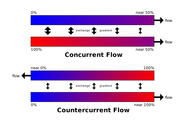
The blood flowing along the gland and the fluid within the gland flow counter, or in opposite directions to one another. One of the principles of osmosis is that molecules will move from fluids with high concentrations of solutes (in this case, salt) to fluids with lower concentrations. I.e. they move down their concentration gradient. So salt leaves blood and goes to the relatively less salty fluid in the duct. Since the blood and the duct fluid are moving in opposite directions, the blood will always remain saltier than the duct fluid, therefore a concentration gradient will be maintained, and the salt will always flow out the blood to the fluid in the duct.

Still confused? There's a pretty great diagram here. For the more verbally inclined, here's a metaphor: Imagine the blood stream and the fluid stream are two trains moving parallel to each other in opposite directions (I promise this won't involve any math). Every car in the "blood" train is full of (rather salty) people, and every car in the fluid train is empty. As the trains meet, the first two cars of each train will be facing each other, and the people from the blood train (with great nimbleness) will jump from the blood train to the fluid train, until the first car in the fluid train is full. Then as the the trains continue to pass, people will continue to jump from the blood train and fill all the cars in the fluid train. People will not jump back onto the blood train from the fluid train because remember, they're going in opposite directions. So as the people that jumped on the first car of the fluid train keep going, they're passing the full cars of the blood train going in the other direction.

What would happen if the blood and the fluid were running in the same direction? That would be called concurrent exchange. The salt would still move from the blood, but quickly the concentrations of salt within the blood and the fluid would become the same and there wouldn't be any net gain or loss of salt in either the blood or the fluid. Pretend those trains are now running in the same direction, and people jump onto the fluid train, but then they see open space in the blood train and jump back. Since there will always be space in both the fluid and the blood trains, and they keep running along next to each other, passengers will keep jumping back and forth.

This diagram illustrates the difference between concurrent (top) and counter-current (bottom) exchange. Blue, in this case, would indicate low salinity, and red would indicate high salinity. As for the percentages, there is never "near 100%" absorption of salt from the bloodstream, as that would be as deadly as having too much salt. So take the percentages with a grain of salt.
From Wikimedia commons


The result is a fluid that is actually saltier than sea water. It flows from the gland and is excreted through the nasal passages. Penguins will often look like they have runny noses, but it's really this salty substance coming out their noses. You could, supposedly, say that they pee out their nose, but that would take away some of the mystique from the magestic emperor penguin, wouldn't it?

Immature jokes aside, this gland allows penguins to consume massive amounts of salt, and still be healthy. Considering how high sodium intake contributes to heart disease, maybe penguins could unlock the biotechnology of the future to allow us to have our salt cake and eat it, too!

Reference:
John Sparks & Tony Soper. (1987). Penguins. Facts on File, Inc. 460 Park Avenue South, New York, NY.

Tuesday, February 28, 2012

Neuromagicology: At the Intersection of Art and Science

We all know how the cameras in our phones are only so good. The photos look grainy and the colors washed out. Compared to the naked eye, phone cameras don't seem to compare.

Well actually, the camera in your smartphone is 2 1/2 times better than your eye! In other words, if the resolution on your phone camera is 5 megapixels, the processing power of your eyes roughly equates to about 2 megapixels. But then, how is it the world around you looks so much sharper, richer, and full of color than the photos on your camera roll? It's because you have something your phone can't even begin to emulate, the brain.

Don't believe me? Hold your arms straight out in front of you. Put the tips of your thumbs together with your index fingers pointed up towards the ceiling, so you're making mirrored L shapes, or one big U shape. Now close your left eye and look at the tip of your left index finger with your right eye. While looking at your left finger tip, focus your attention on your right finger tip. Did it disappear? If it didn't, wiggle it around a bit, and you'll see what I mean; you'll notice that it suddenly vanishes from sight.

That's because your right finger is sitting squarely in the blind spot of your right eye. There are no light-sensing photoreceptors there because that's where all the fibers that make up your optic nerve converge. It has been there all your life, yet you don't notice it until an illusion forces you to. You might have noticed that instead of your finger where it should have been, you just saw the wall or the computer screen or whatever your finger was in front of. What's going on here is the brain is "filling in" that blindspot with the stuff around it. Kind of like the clone stamp tool does in Photoshop.

Illusions reveal the "supreme achievement of the brain"

For a long time, illusions have been thought to be the tools to reveal the limitations of the visual system and show where the brain "got it wrong." Neuroscientists Stephen Macknik and Susana Martinez-Conde see it differently. They think illusions really reveal something special, magical even, about the brain. "This is one of the supreme achievements of the brain," says Stephen, "The brain has actually evolved these processes that are illusory for the purpose of improving vision."

Not only do these illusions show us the nature of our visual experience, but they can also tell us something about consciousness. Consciousness is the first person experience of your life in the world, and it is home grown in your brain. Your senses interact with the outside world and send electrical signals to your brain to make sense of them, but when you look at these sensory systems, "you realize that the information going [to the brain] is really quite deprived." When so little information goes in, the brain has to fill in the details. The so-called conscious part of your brain comes from a separate group of neurons that takes information from your sensory and cognitive systems, your memories, your attention and other systems, and cobbles it together to make a simulation of reality. As Stephen puts it,

That simulation of reality is the only thing you've ever interacted with, it's not that the real world isn't out there--it is--but you've never been there. You've only ever interacted with this simulation of reality that's put together from sparse information from the outside world and the rest is essentially confabulated, just like that blindspot is a confabulation of sorts.

From illusions to magic.

Stephen and Susana are very interested in how our attention and awareness, through the visual system, can be manipulated and what that manipulation says about the process--or confabulation--of consciousness. Illusions can certainly help, but they really only pertain to vision, not awareness and attention in particular. But while organizing a conference for the Association for the Scientific Study of Consciousness in Las Vegas, a little magic happened for Stephen and Susana.

They were brainstorming on how to generate public interest in the topic of consciousness, and they realized that they needed to study the artists of attention and awareness. But who would that be? "Finally it got through to us, Las Vegas spoke to us directly. It said, 'Magicians are the performance artists of attention and awareness.'"

Things took off from there. Stephen and Susana have worked with some great names in magic, like James Randi, Penn & Teller, and Apollo Robbins. Magicians, in the pursuit of bettering their art, have come up with some great theories about how the brain works that neuroscientists have yet to test in the lab. Having these theories before you start to research can also take years off the research process, and really help advance the field of awareness, cognition, and consciousness.

The intersection of art and science

Susana focuses her research on eye movements. There are two different types of eye movements, saccades (which I talked about before) and smooth pursuit. To see the difference. hold out your thumbs in front of you and look at your right thumb. Now try to move your eyes in a line from your right to your left thumb, and you'll notice that you can't do it. Your eyes seem to "skip" along a line to your left thumb. That skipping from point A to point B is called a saccade. Now look at your right thumb as you move slowly to your left, and now you can follow it smoothly, hence "smooth pursuit." So we've just demonstrated to ourselves that smooth pursuit eye movements are involuntary. Now for the Magic.

Apollo Robbins is a professional thief. His act involves very close-range sleight of hand where he pick-pockets from people right before their eyes. Through his art, he noticed that if he moves his hand in a straight line from someone's pocket, people will look at where the hand is going to go, and then immediately back to the pocket through saccadic movement, and that this is a good way to distract someone, or trick them into thinking that he stole something from that pocket when he really didn't. But when he moves his hand in an arch people have to use smooth pursuit to follow his hand, and they don't look back to the pocket at the end.

Apollo's observations led Susana and Stephen to think that perhaps smooth pursuit and saccadic movements affect attention differently, and prompted them to do a study. They found that with straight arm movement away from the pocket from which an item was "stolen," the attention of the thief-ee is directed through saccadic motions from the pocket to Apollo's hand, making the pocket the last place where the thief-ee had their attention, and thus they look back at it. But smooth pursuit eye movement directs attention to the hand as it moves away from the pocket, and there's enough time in between that the pocket isn't the next logical point of attention anymore.

And that isn't the only example, either. Magicians will "use humor in order to, basically, get away with magical murder. If they get people to laugh, their attention is suppressed." When you think about it, this might seem obvious, but there actually isn't any literature in neuroscience on the emotional modulation of attention. Studies on PTSD and anxiety get at the idea, few have looked at the effect of emotions other than fear on attention.

Sleights of Mind

In their book, Sleights of Mind: What The Neuroscience of Magic Reveals About Our Every Day Deceptions, Stephen Macknik and Susana Martinez-Conde explore just that. They look at how magicians intrinsically understand the mechanisms of our attention and awareness and what their manipulation of those mechanisms can tell us about how our brain constructs our sense of reality from sensory stimuli.

Not only is this book very educational, but it's fun! Sleights of Mind is just as much about magic as it is about neuroscience. It's a great read for anyone, regardless of their background in science, who wants to know more about the brain and how it can be hacked. To order the book, and see some really awesome videos, illusions, and more, visit sleightsofmind.com!

Tuesday, February 7, 2012

The Advantage of Being Cute

Most people would agree that babies are pretty damn cute. Put a grown man or woman in a room with an infant, and all bets are off, that baby is getting 100% of that man or woman's attention. Wild horses could not stop most people from cooing a baby, yet we don't usually question why. The truth?

That baby is pretty much helpless on its own, so if it's going to survive, it needs lots of attention from adults. Until it gets to the point where it can walk and talk on its own (and then some), the baby is going to lure you into caring for it with those adorable chubby cheeks and wide eyes full of wonder.

Ethologist Konrad Lorenz first put forth the idea that the typical cute baby, with a large head, round eyes, small nose and mouth, elicits a caregiving response from adults, and even suppresses aggressive behavior.  These features, known as a baby schema or a Kindchenschema, are a pretty useful thing for a baby to have. However, until recently this hypothesis was a bit shaky. Most studies that looked at people's responses to infantile adorableness used line drawings or unmanipulated photos of babies that could not control for other aspects known to affect emotional responses such as facial symmetry.

With the help of Photoshop, researcher Melanie Glocker and her team at the University of Pennsylvania created a situation where participants would only see differences in the baby schema of infants and be able to rate their cuteness and how much they desired to care for the babies. They took pictures of babies and created three photos of each baby: one undoctored, one changed to maximize the baby's cuteness, and one to minimize it.

In the center, the unmanipulated photo. With the less cute manipulated photo to the left, and the cute one to the right.
Then the researchers got a group of 122 undergraduates and split them into two groups: one group would rate each photo's cuteness, and the other would rate how much they wanted take care of the baby in each photo, both on a 1-5 scale.

As expected, participants rated the "high cuteness" manipulated photo as being significantly more cute than both the undoctored and "low cuteness" manipulated photos. Participants in the caregiving group also rated themselves as having a stronger desire to take care of the cuter babies.

Other research has suggested that the emotional impact of cuteness is influenced by female sex hormones, so the researchers hypothesized that women would be more strongly affected than men by cuteness. In fact, men and women rated the cuteness of babies pretty equally, and both men and women had a stronger desire to take care of cuter babies than less cute babies. However, women rated their desire to take care of babies of all cuteness higher than the men did. The researchers suggested that this could be a cultural as well as biological predisposition as historically in many cultures, women have generally been the primary caregivers of children.

The researchers also mentioned that before the rise of the nuclear family, childrearing was often done with the cooperation of friends and extended family. This could explain why both men and women have a strong desire to take care of babies, and why this desire extends to babies that they are not related to. After all, we are social animals, so we have to look out for each other!

Reference:
Glocker, M.L.; Langleben, D.D.; Ruparel, K.; Loughead, J.W.; Gur, R.C.; Sachser, N. (2009). Baby Schema in Infant Faces Induces Cuteness Perception and Motivation for Caretaking in Adults. Ethology 115(3): 257-263.

Special thanks to my wonderful and wonderfully talented friend, Carolyn McGraw, for making that awesome Admiral Ackbar drawing! You can see more of her stuff here and here.

Sunday, January 15, 2012

Blindsight and Consciousness, what can we learn from the blindsighted?

If there were ever a perfect example of an oxymoron, the term blindsight would be it.

Other than the best oxymoron ever, what is blindsight?  Alan Cowey, in his 2010 review article, The blindsight saga, describes it as such:
It is the ability of patients with absolute, clinically established, visual field defects caused by occipital cortical damage to detect, localize, and discriminate visual stimuli despite being phenomenally visually unaware of them.
In simpler terms, it's the ability to sense the presence of objects in one's visual field without consciously seeing them the way normal sighted people do.  While it would be a stretch to call blindsight a superpower, the visual capabilities of a person with blindsight could be considered akin to those of Dare Devil.  Blindsighted people often are able to identify visual stimuli, although they deny having a conscious experience of actually seeing anything.

In this video, a blindsighted patient is able to navigate a field of obstacles successfully, even though he can't see them.

How does blindsight happen?  It is a fairly unique condition; not all people who are blind possess blindsight.  It occurs in patients who become blind in part or all of their visual field after suffering damage to the primary visual cortex, known as V1.
The primary visual cortex (V1) highlighted in yellow.  The bottom view is from a mid-section of the brain, the top view is from the outside.  In both views, your eyes would be on the left.  Source.

Becoming totally "cortically blind," as is the case for the patient in the video above, is actually pretty rare (thankfully), so most patients with blindsight are only blind in part of their visual field, while the rest of  the field remains normally sighted.

A controversial subject

Blindsight has drawn a lot of controversy among researchers.  Since the hallmark trait of blindsight is responsiveness to visual stimuli without the conscious experience of perceiving it, it makes animal studies a little difficult.  You can observe where a cortically blind animal turns its attention when presented a stimulus, but you can't ask it if it actually saw anything.  So researchers interested in getting at the question of what people with blindsight actually do or do not experience have a relatively small pool of subjects they can perform research on.

Then, of course, there's the fact that whatever blindsighted patients report as their experiences of blindsight must be taken with the grain of salt as all experience is subjective.  Some patients report that, even though they didn't see anything, they have a feeling that "something happened."  The question, then is, is this feeling essentially visual in nature?  Did it come about due to light scattering from the stimulus into their seeing field?  Or did the subject report a feeling of something happening because that's what he or she felt that the researchers were looking for?  The answers to these questions have proven to be extremely difficult to tease apart.  Regardless, the fact that some patients have "feelings" when presented a stimulus and others do not requires that blindsight be split into two categories: type 1, no awareness at all; and type 2, awareness without visual experience.

Even in patients with type 2 blindsight, the awareness of the stimuli isn't always consistent.  But we can still learn something here.  In one task, patient GY was asked to discriminate between the presence or absence of a stimulus and then wager money on his answer.  Incorrect wagers would be subtracted from his winnings, thus prompting him to only wager high when he was very confident of his answer.  When he felt that he was aware of the stimulus, despite not seeing anything, he wagered high and was correct more than 90% of the time.  When he didn't report any awareness, he wagered low, as would be expected.  But he was correct more often than could be explained by chance, suggesting that even without awareness, he was still able to perceive visual stimuli.

Total cortical blindness

Let's re-visit the patient in the video, TN.  After successive strokes, TN's primary visual cortex was completely destroyed, as was later verified by both structural and functional MRIs.  Something helpful at least came of TN's misfortune, in that he gave researchers De Gelder et al a rare chance to study a person with total cortical blindness.  The video above is truly astonishing because, at first glance, it looks like he is able to avoid obstacles without the use of a cane or any outside guidance.

Nevertheless, this video is not without its critics.  The first that springs to mind is echolocation, a capability that has been shown in other blind people.*  The researchers recognized that this could not be ruled out, but many critics of the research suggested that not enough attention was paid to the possibility of echolocation anyway.  Other critics say that TN could have been unconsciously processing auditory signals from the researcher shadowing him (to make sure he didn't stumble or fall), and that those aided in his navigation.

TN's contributions to blindsight research don't just end with that video.  TN also demonstrated affective blindsight, or the ability to discriminate between emotional stimuli in a physiological sense.  Researchers Gonzalez-Andino et al showed TN pictures of various facial expressions and monitored his brain activity via an EEG.  Without any awareness of the stimuli whatsoever, the researchers were able to localize changes in the electrical activity of parts of the brain associated with emotional stimuli, such as the right amygdala, in TN.  In these trials, TN was also not asked to guess at the nature of the stimuli, either, so the results suggest some perceptual ability without any conscious awareness at all.

Conclusions: what blindsight can tell us about consciousness

It is extremely important to note that all studies on blindsight and consciousness are done with very small sample sizes.  Most studies are case studies that focus on the abilities of only one patient.  Because of this, everything that we can say about blindsight must be taken with a grain of salt.  There simply aren't enough subjects with blindsight to tell us very much with any certainty about the nature of consciousness and vision.  Nevertheless, some results are so provocative that they can at least give us clues and ideas about where consciousness in vision lies in the brain, and can give us leads for further and more focused studies in the future.

It has become clear that blindsighted patients' vision is certainly unlike normal vision, and obviously unlike total blindness.  There is definitely some ability in patients to perceive objects, even if the true nature of that ability remains murky.  And it's the murkiness of that ability that makes blindsight so tantalizing to researchers interested in the neural mechanisms of consciousness.  How can one see without actually seeing?  That is the big question.  The visual pathway is certainly very complex, but the blindsighted just may be able to tell us what aspects of that pathway give rise to the conscious experience of sight, and wouldn't that be so cool?

*While looking for the video about Ben Underwood, I found out that just a few years after the original news spot was filmed, he sadly died of cancer.  You can read more about him and his life here.

References:
Cowey, A.  (2010).  The blindsight saga.  Experimental Brain Research 200:3-24.
de Gelder, B.; Tamietto, M.; van Boxtel, G.; Goebel, R.; Sahraie, A.; van den Stock, J.; Stienen, B.; Weiskrantz, L.; Pegna, A.  (2008).  Intact navigation skills after bilateral loss of striate cortex.  Current Biology 18,24:1128-1129.
Gonzalez-Andino, S.L.; de Perlata Menendez, R.G.; Khateb, A.; Landis, T.; Pegna, A. (2009). Electrophysiological correlates of affective blindsight.  NeuroImage 44:581-589.

Sunday, December 18, 2011

Can woodpeckers help us design better helmets?

Despite being one of the more annoying animals, woodpeckers are actually pretty cool when you consider how well they're designed for what they do.  Their feet have two toes in front and two in back to better grip vertical surfaces.  Their stiff tails act like a third leg to balance themselves against the tree (or building) that they're pecking.  But what's really cool is their skull.

If you were to sit down and try to design a helmet, you might not think of looking towards nature for inspiration.  But while pecking, a woodpecker's head accelerates and decelerates at very high speeds.  In order to protect itself from injury due to impact, it has evolved several structural mechanisms to absorb the shock of repeatedly ramming its face into a tree.

Some of these structures include a very sharp and stout beak, which minimizes the impact of the beak hitting the tree, much like jumping into water feet-first minimizes the impact of the surface tension on the rest of the body.  The stoutness then helps to absorb most of the shock and prevent it from reaching the cranium of the skull.  There is also less space between the brain and the skull than in other animals, which helps to keep the brain from getting jostled around during impact.  Woodpeckers also have a long hyoid bone, which is attached to the tongue, forks and extends around the neck, up behind the skull, over the top, and into the right nostril, acting as a sling to cradle the skull.
Yes, really.  On the left is the skull of a woodpecker, where the hyoid can be seen extending up from behind the skull.  On the right labeled (b) is the woodpecker hyoid bone by itself.
In a recent article in PLoS ONE, researcher Yubo Fan and co. decided to look more closely at the engineering of the woodpecker skull.  By comparing the structure of a woodpecker to the soil-pecking Eurasian hoopoe using a high-speed videos, force and torque sensors, and micro-CT scanning, they were able to determine how the internal structure of the skull and beak interplay to absorb shock and prevent brain injury in woodpeckers.

First is the composition of the skull itself.  Bones are not entirely solid throughout, they are mostly composed of porous "spongey" bone, with a solid outside.  In the skull of a woodpecker, the spongey bone is made of a dense, plate-like organization, which acts like packing peanuts.  In contrast, the hoopoe has spongey bone that looks more like rods, which is less effective in absorbing shock.

The internal structure of the beak is also specially designed to absorb shock, with a longer outer tissue layer and a very tough internal bone.  In fact, the beak absorbs most of the shock during impact, as shown by this diagram demonstrating the distribution of force (shown in red) throughout the skull over the time of impact and recoil.
Finally, there's the distribution of force on the hyoid bone.  Its unique structure allows it to absorb any extra force from the impact on the beak and divert it away from the skull itself.  It also helps to keep the skull in place, like a seatbelt keeps a person in place during a car crash.

The woodpecker is turning out to be another inspiration from nature to engineers.  When trying to create a better helmet, why not look to the solutions that millions of years of evolution has produced?

Images courtesy of PLoS ONE.
Reference: Wang, L.; Tak-Man Cheung, J.; Pu, F.; Li, D.; Zhang, M.; Fan, Y.  (2011).  Why Do Woodpeckers Resist Head Impact Injury: A Biomechanical Investigation.  PLoS ONE. http://www.plosone.org/article/info%3Adoi%2F10.1371%2Fjournal.pone.0026490

Sunday, December 4, 2011

Home for the holidays! How birds use their sense of smell to find their way home

Happy holidays, everyone! It's a time of eating lots of delicious food, spending time with friends and family, and celebrating long-held traditions. For many, it's also a time of finding their way back home, whether it's in the town where they grew up, or in the company of loved ones (or both). This also means that for many, it's a time of airports and cars and lots of frustrating travel. For us humans, navigating home involves making reservations, getting on a plane in one city and landing in another. Or it means climbing into the car, punching in an address in the GPS, and hitting the gas. But what does getting home mean for other animals? They don't have a GPS with a vaguely snarky voice to tell them which way to turn, nor do they have massive(ly disorganized) transportation hubs in major cities that quickly shuttle them back and forth to destinations. So what happens when you take an animal, put it somewhere where it's never been, and let it try and find its way home?

That's actually a pretty big question when it comes to animal navigation. Different animals have very different ways to navigate--for example, some use the position of the sun to orient themselves. Others can see polarized light, and use that to navigate home. Yet others use strategies more familiar to us, they look for familiar visual cues and landmarks, and use that to orient themselves. You've also probably heard of animals using magnetic fields (known as magnetoception) to discern their location on the globe. Most famous for this form of navigation are the birds, who have small particles of magnetite in their upper beak that allow them to sense the magnetic fields over the earth, and orient themselves within those fields. This form of navigation comes in handy quite a bit, especially on overcast days when birds cannot use a sun compass effectively, or when they're in an unfamiliar location and cannot identify any familiar landmarks. Magnetoception certainly plays a very important role in bird migration but, surprisingly, may not be the go-to strategy for a bird finding its way back to a familiar "home base" from a completely novel location. In this situation, studies have shown that it may be a bird's sense of smell that it primarily relies on to find its way back home.

Homing pigeons are most commonly used as model species to determine the homing ability of birds. However, determining exactly how they navigate is a bit tricky. They have an innate ability to use magnetoception (meaning they're born with the ability), but they have to learn to use the magnetic compass in relation with familiar visual landmarks to get a sense of a "magnetic map" in order to find their way home. When they're about three months old, they then use to learn a solar compass to aid in navigation, by using the position of the sun in relation to the magnetic and visual cues. Most studies use young birds that haven't learned the sun compass yet, or birds that haven't been allowed to learn a visual map (by keeping them in captivity), to determine innate homing capabilities. When you bring these birds to a novel location and let them go, they are usually able to fly home, even if you prevent them from using the magnetic compass by severing the nerve that relays the magnetic information to the brain. However, if you take these same birds and make them anosmic (unable to smell), they tend to get lost and have a hard time finding their way home, if they do at all. But what happens when you take adult birds that have been allowed to learn a visual map and put them in the same conditions?

A study by Gagliardo et al in 2009 did just that. They allowed birds to become familiar with the area around their home loft, and then split them up into three groups. One group had the opthalamic branch of the trigeminal nerve--which relays magnetic information to the brain--severed, rendering their magnetic compass ineffective. Another group had their olfactory nerves severed, rendering them anosmic. And another group was left intact as a control. They then took these birds to novel locations either 50-60 km, or 75-100km away from their home roost, let them go, and observed their homing abilities. Here are their results:

This is a figure from the study representing the homing success of the birds at locations 50-60km away from the home loft. The three circles at the top of the figure represent the three groups, with C being the control, V1 being the magnetically-impaired group, and ON being the olfactory (smell)-impaired group. The "H" at the top of the circles represents the direction of the home loft from the release site, and the arrow from the center represents the average vector of the direction the birds were flying in when they vanished from sight. Each triangle represents the direction of the individual birds of each group as they vanished from sight. In both the control and magnetically-impaired groups, the birds were generally flying in the direction of the home loft, but in the olfactory-impaired group, they were generally flying away from home.

The bottom-left section shows the homing speed (in km/hr) of the pigeons that found their way home the same day of the release. To the right shows the number of pigeons (represented again by each triangle representing an individual pigeon) that found their way home the next day, more than one day later, and ones that never returned home, or were "lost." This shows a pretty clear demonstration that when pigeons were deprived of their sense of smell, they were more likely to become completely disoriented and lost after being released from a novel location. The data from the group released from 75-100 km from home shows a similar pattern, though all the pigeons in each group showed greater difficulty in homing regardless of their sensory capabilities.

Whether or not the magnetic compass or olfaction is the primary navigational tool for homing in birds is still pretty heavily debated. It's been suggested that the various methods to deprive birds of smell may also get in the way of magentoception, because the nerves and sensory organs involved in both senses are in very close proximity to each other in the birds' anatomy. However, this and other studies provide quite a bit of evidence that olfaction is very important in a bird's ability to find its way home. This study in particular lends a lot of weight to the idea that olfaction is necessary for navigation considering it used adult pigeons that were experienced in navigation, unlike many studies of the past.

So, if you have the misfortune of getting stranded in an airport or stuck in a traffic jam while making your way home this holiday season, you can at least take some comfort that you don't have to rely on your sense of smell to get home!

Reference: Gagliardo, A.; Paolo, I.; Savani, M.; Wild, M. (2009). Navigational abilities of adult and experienced homing pigeons deprived of olfactory or trigeminally mediated magnetic information. The Journal of Experimental Biology 212, 3119-3124.

Thursday, November 17, 2011

Gender-Bending in the Animal Kingdom

An article in the New York Times this week inspired me to write about my favorite animal of all time, the cuttlefish.

:D


Cuttlefish are cousins of squids and octopuses.  They're not native to the Americas (which is why many American readers may have never heard of them before), but they are common virtually everywhere else on the globe.  Like their cousins, cuttlefish are capable of extremely elaborate camouflage, a skill which can be and is used for hunting and communication, as well.  They're like chameleons, only about 10,000x cooler.  Why?

That's why.

Cuttlefish (and other cephalopods) have specialized pigment cells called chromatophores.  In the cuttlefish, these chromatophores are like little sacks of color that come in brown, yellow, and red.  Other animals, like chameleons, also have chromatophores, but what makes cephalopod chromatophores so unique is that they are surrounded by muscles, which are innervated by nerves.  Here's a fairly simple diagram:
When those muscles around the chromatophore contract, it expands the radius of the pigment cell, creating a visible dot of color, much like a pixel on a screen.  Cuttlefish skin is full of millions of these cells.  But the awesomeness doesn't end there.  Underneath the layers of chromatophores are colorless, light-reflecting cells called iridophores and leucophores, which reflect green, blue, pink, orange, and white.  The combination of all these various pigment and reflecting cells, along with the papillae that allow cuttlefish to change the texture of their skin by forming little bumps, creates a complete palette that allows the cuttlefish to blend into virtually any environment.  To demonstrate the point, here's a video of an octopus using the same type of skin to blend into its environment.


This complex camouflage is a rather useful strategy for cephalopods.  With the exception of the even less famous, but far more threatened Nautilus, cephalopods have very little protection from predators.  Their ancestors secreted large, thick shells that protected them, but squids have an extremely reduced internal shell (called a pen), and octopuses have no shell at all.  Cuttlefish have an internal porous shell called a cuttlebone, put even that doesn't offer much protection.  These animals are basically soft sacks of protein that everything (including humans) wants to eat.  While all cephalopods can secrete ink to make a quick escape from a predator, the camoflauge helps them to avoid being seen by predators in the first place.

Of course the byproduct of having millions of cells surrounded by muscles which are controlled by nerves is a big brain.  Perhaps what makes cephalopods so famous and charismatic is their intelligence.  You often hear stories about researchers and aquariums losing track of octopuses because they figured out how to escape from their tanks.  Cephalopods have also been shown to be able to learn visual cues to help solve mazes, and even use tools.

The trademark cephalopod cleverness brings me back to the original reason I wrote this post (aside from my fanatic love of cephalopods and talking about them).  One of the most amazing demonstrations of camouflage and intelligence in cuttlefish is seen in the case of "cross-dressing" in Australian Giant cuttlefishes, Sepia apama.  These cuttlefish are solitary, except for once a year where they have a mass mating frenzy.  During this time, the males who would usually like to go unnoticed put on these vibrant displays of colors and patterns to attract the females.  However, the females get the final choice in the mating game.  The way cuttlefish mating works is the male cuttlefish places a packet of sperm on the female's underside, which she later uses to fertilize her eggs.  Females will mate with a few males but they are also very picky about who even gets to mate, rejecting 70% of mating attempts.

Usually, it's the biggest, brawniest, most colorful cuttlefish that gets to mate with the female and later gets chosen to fertilize her eggs.  In part that's because he's big enough to be able to guard her and prevent other males from moving in.  Smaller males barely stand a chance against the largest, guarder males, so some of them employ a clever trick to get past their burly competitors by cross-dressing!  They assume the color pattern of the female and hide their uniquely masculine arms by curling them under, and then they just simply swim past the bigger male virtually unnoticed.  Unless, of course, it's so convincing that sometimes males will try and mate with these so-called sneaker males.  But the more important question is what do the ladies think?  Dr. Roger Hanlon looked into this question by subjecting the cuttlefish to paternity tests.  It turns out that when choosing who gets to fertilize her eggs, the female will select the sneaker males second to the big, brawny males, as if to give an "evolutionary nod" to the cleverness of the sneaker males.  That's not to say that she's consciously acknowledging the clever strategy (consciousness in animals is an entirely different discussion all together), but obviously there is an advantage to having this level of intelligence among cuttlefish, as it gets passed on to the next generation fairly reliably.

Who says the jocks get all the chicks?

Information from NOVA's Cuttlefish: Kings of Camouflage and Hanlon, RT; Naud, MJ; Shaw, PW; Havenhand, JN.  (2005).  Nature 433(7023) 212-212.


*Edit* It was pointed out to me that I referred to iridophores and leucophores as pigment cells, which is misleading as these cells do not actually contain pigment.  They are colorless but reflect certain wavelengths.  Leucophores reflect white light, whereas iridophores reflect pink, orange, green, and blue.